胶原诱导性关节炎小鼠(CIA)作为人类类风湿关节炎模型应用广泛,但CIA引起的关节炎起病比较缓慢,造模周期较长,一般为6-8周(1-12)。Chondrex公司已开发出单一种单克隆抗体合剂诱导的小鼠关节炎模型(CAIA),明显缩短了造模周期。与CIA相比,CAIA模型具有以下优点: (1)可在大多数小鼠上诱发关节炎,包括CIA不敏感的小鼠;(2)CAIA造模周期短,加速筛选和评估类风湿性关节炎治疗药物;(3)应用于转基因小鼠来研究基因对关节炎发病的影响;(4)用于研究与人类类风湿性关节炎相关的各种炎症介质和因素在疾病中的作用,例如细菌和病毒毒素(13-17)。
CAIA without LPS
胶原抗体诱导的关节炎(不加LPS)
CIA是由于自身抗体形成的免疫复合物通过激活补体而诱发关节炎症,因此研究表明多克隆胶原蛋白抗体(8-19) 或单克隆胶原蛋白抗体合剂能诱发关节炎(20-21)。根据MHC类型,这些自身反应性抗体可以识别位于小鼠II型胶原蛋白的CB11或者CB8片段中的特定抗原定簇(关节炎抗原表位)(22-23)。
Chondrex公司生产的能诱发小鼠关节炎的抗体合剂含有 : A2-10 (IgG2a), F10-21 (IgG2a), D8-6 (IgG2a), D1-2G(IgG2b), and D2-112 (IgG2b). 其中两个单克隆抗体(F10-21和D8-6)识别在LysC-2(291-374)的83个氨基酸肽段,该肽段有LysC内切酶作用产生。 另外三个单克隆抗体(A2-10,D1-2G和D2-112) 能够识别二型胶原蛋白CB11片段(124-402)中LysC-1(124-290)的167各氨基酸肽段(13)。这些抗原表位氨基酸序列在各种物种中具有高度保守性,包括鸡,小鼠,大鼠,牛,猪,猴子和人 (20-21)。
CAIA with LPS
胶原抗体诱导的关节炎(加LPS)
研究表明,联合注射低于致炎剂量的单克隆抗体合剂和大肠杆菌多脂(LPS)可诱发严重的小鼠关节炎(13)。通过胃肠道吸收的细菌毒素(如LPS)与低致炎水平的二型胶原自身抗体对引发关节炎有发协同作用(24)。这种模型相比于传统CIA 模型有多重优势。第一,起病时间短,发病率高达100%。第二,可在大多数小鼠上诱导关节炎,包括CIA不敏感的小鼠,T细胞缺陷的小鼠,特定基因敲除变异的小鼠,详情请见表二.
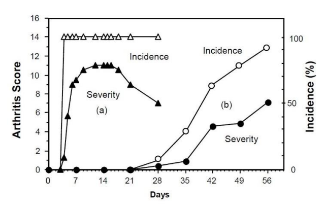
图一显示CIA模型和CAIA模型特点的比较。CAIA造模周期是CIA建模周期的十分之一。

图一 CAIA vs. CIA: (a)三角形:100%的小鼠在LPS注射后24-48小时即可见关节炎发作,在第5-7天病情达到高峰。(b)圆形:CIA诱发小鼠关节炎模型一般需要4周起病,即使使用CIA易感品系,如DBA/1J和B10.RIII小鼠。
表二 关节炎炎症程度的临床评分
| 得分 | 发病情况 |
| 0 | Normal 正常 |
| 1 | Mild, but definite redness and swelling of the ankle or
wrist, or apparent redness and swelling limited to
individual digits, regardless of the number of affected
digits
轻度的、踝关节、腕关节发红、肿胀 |
| 2 | Moderate redness and swelling of ankle or wrist
踝关节或腕关节中度发红肿胀 |
| 3 | Severe redness and swelling of the entire paw
including digits
爪子严重发红、肿胀,包括指端 |
| 4 | Maximally inflamed limb involving multiple joints
四肢最大程度发炎,包括多关节 |
引用文献:
1. E. Trentham, A. Townes, A. Kang, Autoimmunity to Type II Collagen an Experimental Model of Arthritis. J Exp Med. 146,857-68 (1977).
2. J. Courtenay, M. Dallman, A. Dayan, A. Martin, B. Mosedale,Immunisation Against Heterologous Type II Collagen Induces Arthritis in Mice. Nature. 283, 666-8 (1980).
3. S. Cathcart, K. C. Hayes, W. A. Gonnerman, A. A. Lazzari,C. Franzblau, Experimental Arthritis in a Nonhuman Primate. I.Induction by Bovine Type II Collagen. Lab Invest. 54, 26-31(1986).
4. P. Wooley, Collagen-induced arthritis in the mouse.Methods Enzymol 162, 361-373 (1988).
5. P. Wooley, H. Luthra, M. Griffiths, J. Stuart, A. Huse, C.David, et al., Type II Collagen-Induced Arthritis in Mice. IV.Variations in Immunogenetic Regulation Provide Evidence for Multiple Arthritogenic Epitopes on the Collagen Molecule. JImmunol. 135, 2443-51 (1985).
6. R. Holmdahl, L. Jansson, E. Larsson, K. Rubin, L.Klareskog, Homologous Type II Collagen Induces Chronic and Progressive Arthritis in Mice. Arthritis Rheum. 29, 106-13 (1986).
7. R. Ortmann, E. Shevach, Susceptibility to Collagen-Induced Arthritis: Cytokine-Mediated Regulation. Clin Immunol. 98, 109-18 (2001).
8. W. C. Watson, A. S. Townes, Genetic Susceptibility to Murine Collagen II Autoimmune Arthritis. Proposed Relationship to the IgG2 Autoantibody Subclass Response, Complement C5,Major Histocompatibility Complex (MHC) and non-MHC Loci. JExp Med. 162, 1878-91 (1985).
9. K. Campbell, J. A. Hamilton, I. P. Wicks, Collagen-induced Arthritis in C57BL/6 (H-2b) Mice: New Insights Into an Important Disease Model of Rheumatoid Arthritis. Eur J Immunol. 30, 1568-75 (2000).
10. E. Michaëlsson, V. Malmström, S. Reis, A. Engström, H.Burkhardt, R. Holmdahl, et al., T Cell Recognition of Carbohydrates on Type II Collagen. J Exp Med 180, 745-9(1994).
11. M. Andersson, R. Holmdahl, Analysis of Type II CollagenReactive T Cells in the Mouse. I. Different Regulation of Autoreactive vs. Non-Autoreactive Anti-Type II Collagen T Cells in the DBA/1 Mouse. Eur J Immunol 20, 1061-6 (1990).
12. R. Reife, N. Loutis, W. Watson, K. Hasty, J. Stuart, SWRMice Are Resistant to Collagen-Induced Arthritis but Produce Potentially Arthritogenic Antibodies. Arthritis Rheum. 34, 776-81
(1991).
13. K. Terato, D. Harper, M. Griffiths, D. Hasty, X. Ye, et al.,Collagen-induced Arthritis in Mice: Synergistic Effect of E. ColiLipopolysaccharide Bypasses Epitope Specificity in the Induction
of Arthritis With Monoclonal Antibodies to Type II Collagen.Autoimmunity 22, 137-47 (1995).
14. S. Yoshino, E. Sasatomi, Y. Mori, M. Sagai, Oral Administration of Lipopolysaccharide Exacerbates CollagenInduced Arthritis in Mice. J Immunol. 163, 3417-22 (1999).
15. B. Cole, M. Griffiths, Triggering and Exacerbation of Autoimmune Arthritis by the Mycoplasma Arthritidis Superantigen MAM. Arthritis Rheum. 36, 994-1002 (1993).
16. Y. Takaoka, H. Nagai, M. Tanahashi, K. Kawada,Cyclosporin A and FK-506 Inhibit Development of SuperantigenPotentiated Collagen-Induced Arthritis in Mice. Gen Pharmacol.30, 777-82 (1998).
17. T. Kagari, H. Doi and T. Shimozato. The importance of IL-1β and TNF-α, and the noninvolvement of IL-6, in the development of monoclonal antibody-induced arthritis. J Immunol 169,1459-1466 (2002).
20. K. Terato, K. Hasty, R. Reife, M. Cremer, A. Kang, J. Stuart,et al., Induction of Arthritis With Monoclonal Antibodies to Collagen. J Immunol 148, 2103-8 (1992).
21. P. Hutamekalin, T. Saito, K. Yamaki, N. Mizutani, D. Brand,et al., Collagen Antibody-Induced Arthritis in Mice: Development of a New Arthritogenic 5-clone Cocktail of Monoclonal Anti-Type II Collagen Antibodies. J Immunol Methods 343, 49-55 (2009).
22. K. Terato, K. A. Hasty, M. A. Cremer, J. M. Stuart, A. S.Townes, A. H. Kang, et al., Collagen-induced Arthritis in Mice.Localization of an Arthritogenic Determinant to a Fragment of the Type II Collagen Molecule. J Exp Med. 162, 637-46 (1985).
23. L. Myers, H. Miyahara, K. Terato, J. Seyer, A. Kang,Collagen-induced arthritis in B10.RIII mice (H-2r): identification of an arthritogenic T cell determinant. Immunol 84, 509-513 (1995).
更多资料或相关产品,请联系Chondrex全国授权代理-欣博盛生物
全国服务热线: 4006-800-892 邮箱: market@neobioscience.com
深圳: 0755-26755892 北京: 010-88594029
广州:020-87615159 上海: 021-34613729
代理品牌网站: www.neobioscience.com
自主品牌网站: www.neobioscience.net